Magnetic Resonance Imaging: Principles, Sequences, and Clinical Applications
Magnetic resonance imaging (MRI), also known as nuclear magnetic resonance imaging (NMRI) or magnetic resonance tomography (MRT), is a sophisticated medical imaging technique used to visualize the internal structures of the body. Unlike X-ray-based methods, MRI relies on the magnetic properties of atoms—primarily hydrogen protons—to create highly detailed images of soft tissues, organs, and neurological pathways.
By utilizing powerful magnetic fields and radiofrequency pulses, MRI allows clinicians to distinguish between different types of biological tissues with remarkable precision. This capability makes it an indispensable tool in modern diagnostics, ranging from neuroimaging and cardiovascular assessment to musculoskeletal and gastrointestinal studies.
Key Facts

- Core Mechanism: Uses strong magnetic fields and radiofrequency pulses to manipulate proton spins.
- Primary Contrast: T1 and T2 relaxation times are the fundamental drivers of image contrast.
- Versatility: Capable of specialized imaging including angiography, spectroscopy, and functional MRI (fMRI).
- Safety Note: Due to the powerful magnets, external metallic objects must be strictly controlled near the scanner.
The Physics of MRI Construction
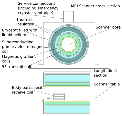
A standard MRI scanner is a complex piece of engineering designed to create a controlled magnetic environment. The system typically consists of a cylindrical superconducting magnet. Within this cylinder, several critical components work in unison:
- Primary Coil: Generates the main, uniform magnetic field.
- Gradient Coils: Create controlled variations in the magnetic field to allow for spatial encoding (locating where signals come from in the body).
- RF Transmit and Receive Coils: Transmit radiofrequency pulses to excite protons and receive the resulting signals.
Signal and Relaxation: T1 and T2
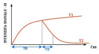
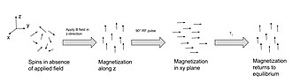
The diagnostic power of MRI stems from how different tissues respond to magnetic pulses. This response is characterized by two main relaxation processes: T1 relaxation (spin-lattice) and T2 relaxation (spin-spin).
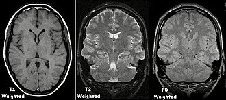
T1-weighted imaging is excellent for visualizing anatomy. In these scans, fat appears with a high signal (bright), while water-rich structures like cerebrospinal fluid (CSF) appear dark. This makes it useful for identifying fat-rich tissues and certain paramagnetic substances like gadolinium.
T2-weighted imaging is highly sensitive to water content. In T2 scans, tissues with high water content—such as those involved in edema (swelling), tumors, inflammation, or infection—appear bright. This makes T2 a primary tool for detecting pathology.
Common MRI Sequences and Clinical Uses
Radiologists select specific "sequences" depending on the clinical question. A sequence is a programmed set of pulses designed to highlight specific tissue characteristics.
| Sequence Group | Primary Physics | Clinical Distinction/Example |
|---|---|---|
| Spin Echo (T1) | T1 relaxation | High signal for fat and contrast agents; anatomical foundation. |
| Spin Echo (T2) | T2 relaxation | High signal for water/edema; detects inflammation. |
| Proton Density (PD) | Proton density | High signal for meniscus tears in joints. |
| Diffusion Weighted (DWI) | Water molecule diffusion | Evaluating cellularity and acute ischemia. |
| FLAIR | Fluid suppression | Nulls CSF signal to better see lesions near fluid. |
| STIR | Fat suppression | High signal for edema in bone or soft tissue. |
Advanced Imaging Modalities
Beyond standard anatomical scans, MRI can be adapted for highly specialized functions:
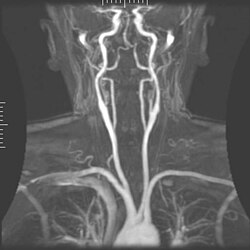
- Magnetic Resonance Angiography (MRA): Visualizes blood vessels to detect aneurysms, stenosis (narrowing), or dissections.
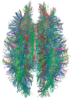
- Diffusion Tensor Imaging (DTI): Maps white matter tracts in the brain to evaluate deformation caused by tumors.
- Functional MRI (fMRI): Uses Blood-Oxygen-Level Dependent (BOLD) imaging to map brain activity.
- Magnetic Resonance Spectroscopy (MRS): Analyzes chemical metabolites within tissues.
Patient Experience and Artifacts
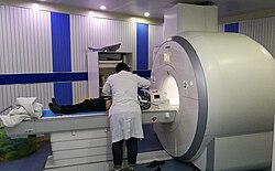
During an MRI, the patient is positioned within the scanner, often using specialized coils to optimize signal for specific body parts, such as the head or abdomen.
While MRI provides exceptional detail, images can sometimes be obscured by artifacts. These are errors in the image that do not represent actual anatomy. Common causes include patient motion, which can blur the image, or susceptibility artifacts caused by metal or certain substances in the body.
![Motion artifact (T1 coronal study of cervical vertebrae)[149]](/images/37/f3/37f3ebd8000664fc53a81c825291e56b79b70327ff5004f9816b025b81fbd09b.jpg)
Frequently Asked Questions
What is the difference between T1 and T2 weighted images?
T1-weighted images are generally better for looking at anatomy, where fat appears bright and water appears dark. T2-weighted images are better for looking at pathology, as they make water and edema (swelling) appear bright.
Is MRI safe?
MRI is generally considered safe as it does not use ionizing radiation (unlike X-rays or CT scans). However, the extremely strong magnetic field poses risks if metallic objects or certain medical implants are brought into the scanning room.
What are contrast agents in MRI?
Contrast agents, such as gadolinium, are substances injected into the patient to enhance the visibility of specific structures, such as blood vessels or tumors, by altering the local magnetic properties.
Why do I need to stay still during an MRI?
Movement during the scan can cause motion artifacts, which result in blurred or distorted images, potentially making it difficult for the radiologist to provide an accurate diagnosis.
What is Diffusion Weighted Imaging (DWI)?
DWI measures the random motion (diffusion) of water molecules within tissues. This is particularly useful in detecting acute strokes or characterizing certain types of tumors.