cyanosiscentral cyanosisperipheral cyanosisdeoxyhemoglobinhypoxia

Cyanosis: Causes, Types, and Clinical Recognition

Cyanosis: Recognizing the Bluish Discoloration of Tissues Cyanosis is a clinical sign characterized by a bluish-purple discoloration of the skin and mucous membranes. This change in color...

Cyanosis: Recognizing the Bluish Discoloration of Tissues

Cyanosis is a clinical sign characterized by a bluish-purple discoloration of the skin and mucous membranes. This change in color occurs when there is a decrease in the amount of oxygen bound to the hemoglobin—the protein in red blood cells responsible for carrying oxygen—within the capillary beds. Because it is a visible indicator of underlying physiological issues, cyanosis is a critical sign for medical professionals in fields such as pulmonology, cardiology, and emergency medicine.

While the term may sound alarming, it is important to understand that cyanosis is not a disease itself, but rather a symptom of an underlying condition. It is most easily observed in areas where the skin is thin, such as the lips, nail beds, ear lobes, and mucous membranes.
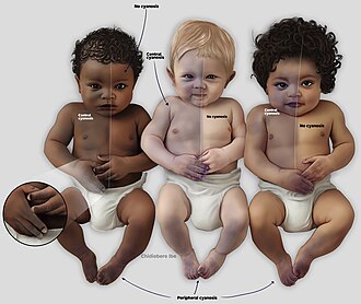
Presentation of cyanosis varies depending on the color of the skin
: Presentation of cyanosis varies depending on the color of the skin

Key Facts

  • Definition: A bluish-purple skin tint caused by low oxygen levels in the blood.
  • Primary Mechanism: An increase in deoxyhemoglobin (hemoglobin without oxygen).
  • Main Types: Central cyanosis and peripheral cyanosis.
  • Critical Threshold: Central cyanosis often appears when oxygen saturation (SaO2) drops below 85%.
  • Diagnosis: Requires physical examination and may involve pulse oximetry, blood gases, or imaging.

Understanding the Types of Cyanosis

Medical professionals classify cyanosis into two primary categories based on the underlying mechanism: central and peripheral.

Central Cyanosis

Central cyanosis is caused by a decrease in arterial oxygen saturation. This typically indicates a systemic issue involving the respiratory or circulatory systems. It becomes clinically apparent when the concentration of deoxyhemoglobin reaches at least 5.0 g/dL (or an oxygen saturation of ≤ 85%).

Common causes of central cyanosis include:

  • Respiratory issues: Pneumonia, asthma (bronchospasm), COPD, pulmonary embolism, or choking.
  • Cardiovascular issues: Congenital heart disease (such as Tetralogy of Fallot), heart failure, or myocardial infarction.
  • Central Nervous System issues: Intracranial hemorrhage, drug overdoses, or seizures that impair breathing.
  • Other factors: High altitude, hypothermia, or rare hemoglobin disorders like methemoglobinemia.
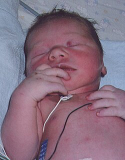
A baby with a heart condition. Note purple nailbeds.
: A baby with a heart condition. Note purple nailbeds.
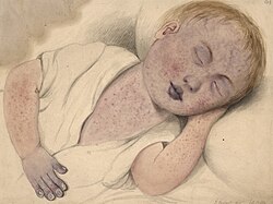
Child with congenital heart disease with central cyanosis that is worsened by measles. Note the bluish-purple discoloration of the fingernails, lips, eyelids, and nose, along with prominent nail clubbing.
: Child with congenital heart disease with central cyanosis that is worsened by measles. Note the bluish-purple discoloration of the fingernails, lips, eyelids, and nose, along with prominent nail clubbing.

Peripheral Cyanosis

Peripheral cyanosis occurs when there is an increased concentration of deoxyhemoglobin specifically on the venous side of the peripheral circulation. Unlike central cyanosis, this is often localized to the extremities, such as the fingers or toes, and can occur even if the heart and lungs are functioning normally.

Common causes include:

  • Reduced cardiac output: Such as in cases of hypovolemia.
  • Temperature extremes: Exposure to extreme cold.
  • Circulatory obstructions: Peripheral vascular disease, Raynaud phenomenon, or deep vein thrombosis (venous obstruction).
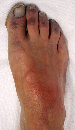
Peripheral cyanosis in an individual with peripheral vascular disease.
: Peripheral cyanosis in an individual with peripheral vascular disease.
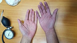
This illustration depicts a self-induced local (tissue) hypoxia on the right hand (right side of the picture) versus a normal left hand (left side of the picture). The cyanosis was achieved by inflating and tightening the blood pressure cuff on the right arm.
: This illustration depicts a self-induced local (tissue) hypoxia on the right hand (right side of the picture) versus a normal left hand (left side of the picture). The cyanosis was achieved by inflating and tightening the blood pressure cuff on the right arm.
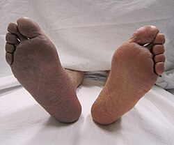
Cyanosis of the right lower limb due to arterial thrombosis (on the left side of the picture).
: Cyanosis of the right lower limb due to arterial thrombosis (on the left side of the picture).

Differential Cyanosis

A specific variation known as differential cyanosis occurs when the lower extremities and abdomen appear bluish, while the head and upper extremities remain pink. This is a hallmark of a patent ductus arteriosus (a persistent connection between the aorta and pulmonary artery). As pulmonary pressure increases, blood flow reverses, sending deoxygenated blood into the descending aorta while sparing the vessels supplying the upper body.
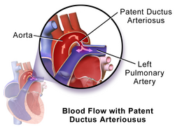
Initial direction of blood flow in patients with patent ductus arteriosus. Once the pressure of the pulmonary arteries increases more than the aorta due to right heart hypertrophy, the direction of blood flow reverses, sending deoxygenated blood through the patent duct directly into the descending aorta while sparing the brachiocephalic trunk, left common carotid, and left subclavian artery, therefore causing the differential cyanosis.
: Initial direction of blood flow in patients with patent ductus arteriosus. Once the pressure of the pulmonary arteries increases more than the aorta due to right heart hypertrophy, the direction of blood flow reverses, sending deoxygenated blood through the patent duct directly into the descending aorta while sparing the brachiocephalic trunk, left common carotid, and left subclavian artery, therefore causing the differential cyanosis.

Clinical Evaluation and Diagnosis

Diagnosing the cause of cyanosis requires a thorough medical history and physical examination. Because skin pigmentation can affect visibility, clinicians must pay close attention to the tongue, mucous membranes, and nail beds, where the skin is thinner.

In newborns, peripheral cyanosis typically affects the distal extremities and the area around the mouth and eyes, but the mucous membranes will remain pink. In contrast, central cyanosis will cause the mucous membranes to appear blue.

Diagnostic tools may include:

  • Pulse oximetry and arterial blood gas tests.
  • Complete blood counts and methemoglobin levels.
  • Electrocardiograms (ECG) and echocardiograms.
  • Imaging such as X-rays or CT scans.
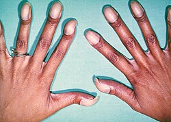
An example of cyanosis in an individual with darker skin pigmentation. Note the pale purple (instead of the typical bluish-purple hue) nail beds. This patient also had prominent digital clubbing due to a congenital heart disease with right-to-left shunting (this patient had Tetralogy of Fallot).
: An example of cyanosis in an individual with darker skin pigmentation. Note the pale purple (instead of the typical bluish-purple hue) nail beds. This patient also had prominent digital clubbing due to a congenital heart disease with right-to-left shunting (this patient had Tetralogy of Fallot).
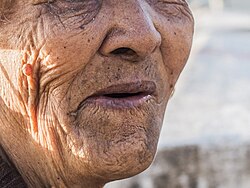
An example of cyanosis in an elderly individual with darker skin pigmentation. Note the dark purple hue of the lips.
: An example of cyanosis in an elderly individual with darker skin pigmentation. Note the dark purple hue of the lips.

Summary of Cyanosis Types

Comparison of Central and Peripheral Cyanosis
Feature Central Cyanosis Peripheral Cyanosis
Primary Cause Low arterial oxygen saturation Increased deoxyhemoglobin in peripheral veins
Mucous Membranes Cyanotic (blue) Pink (normal)
Common Locations Lips, tongue, mucous membranes Fingers, toes, extremities
Typical Underlying Issues Lung or heart disease Cold exposure or local circulation issues

Frequently Asked Questions

Is cyanosis a disease?

No, cyanosis is a symptom of an underlying medical condition, such as respiratory distress, heart problems, or exposure to extreme cold. Management focuses on treating the root cause.

How does skin color affect the diagnosis?

Cyanosis can be more difficult to detect in individuals with darker skin pigmentation. Clinicians look for discoloration in areas with thinner skin and higher vascularity, such as the nail beds, tongue, and mucous membranes.

What are the common symptoms associated with cyanosis?

In addition to the bluish skin tint, associated symptoms may include difficulty breathing, coughing, wheezing, numbness in the affected area, and hypothermia.

Can certain foods or medications cause skin discoloration?

Yes. The consumption of foods with blue or purple dyes, certain birthmarks (like Mongolian spots), and medications containing silver or amiodarone can cause skin discoloration that may be mistaken for cyanosis.

What should be done in an emergency involving cyanosis?

In emergency situations, medical priority is to secure the patient's airway, breathing, and circulation. Supplemental oxygen is often administered immediately for those in respiratory distress.

References

  1. Note this causes "spurious" cyanosis, in that, since methemoglobin appears blue, the patient can appear cyanosed even in the presence of a normal arterial oxygen level.
  2. Note a rare condition in which there is excess sulfhemoglobin (SulfHb) in the blood. The pigment is a greenish derivative of hemoglobin which cannot be converted back to normal, functional hemoglobin. It causes cyanosis even at low blood levels.
  3. Adeyinka, Adebayo; Kondamudi, Noah P. (2021), "Cyanosis", StatPearls, Treasure Island (FL): StatPearls Publishing, PMID 29489181, retrieved 2021-10-28
  4. Dereure, Olivier (2001). "Drug-Induced Skin Pigmentation: Epidemiology, Diagnosis and Treatment". American Journal of Clinical Dermatology. 2 (4): 253–262. doi:10.2165/00128071-200102040-00006. ISSN 1175-0561. PMID 11705252. S2CID 22892985.
  5. Conlon, Joseph D; Drolet, Beth A (2004). "Skin lesions in the neonate". Pediatric Clinics of North America. 51 (4): 863–888. doi:10.1016/j.pcl.2004.03.015. PMID 15275979.