Congenital Melanocytic Nevus: Characteristics and Clinical Signs
A congenital melanocytic nevus is a pigmented skin lesion present at birth. These lesions typically appear as circumscribed patches or plaques, ranging in color from light brown to black. Because they can vary significantly in consistency and size, they may cover any part of the body and any surface area.
Unlike common acquired moles, these nevi are often larger in diameter and may exhibit distinct physical characteristics as the individual grows.
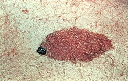
Key Facts
- Appearance: Ranges from light brown to black patches or plaques.
- Growth: Typically grows proportionally with the child's body size.
- Hypertrichosis: The presence of excess terminal hair within the lesion.
- Giant Nevi: Defined by a projected adult diameter exceeding 40 cm (16 in).
- Prevalence: The largest forms occur in approximately 0.002% of births.
- Associated Risks: Potential link to neurocutaneous melanosis in some patients.
Classification and Physical Evolution
Congenital melanocytic nevi are categorized primarily by their size and the presence of hair. A common feature is hypertrichosis, which refers to the growth of excess terminal hair. When a nevus has a projected adult diameter of more than 40 cm (16 in) and exhibits hypertrichosis, it may be referred to as a giant hairy nevus, though the broader term large or giant congenital melanocytic nevus is more frequently used.
Development Over Time
As a child matures, the nevus generally grows in proportion to their body. While some lesions are elevated from birth, many develop thickness and become raised over time. The appearance often shifts with maturity, characterized by:
- Increased development of prominent terminal hairs, particularly after puberty.
- Variations in skin coloration.
- The emergence of textured surfaces featuring proliferative growths.
Neurocutaneous Melanosis
In some cases, these skin lesions are associated with neurocutaneous melanosis, a condition characterized by the coexistence of congenital melanocytic nevi on the skin and melanocytic tumors in the leptomeninges (the membranes surrounding the brain and spinal cord) of the central nervous system.
This association is most notable in those with giant congenital melanocytic nevi, though it can occur with non-giant nevi as well. It is estimated that between 2% and 45% of patients with giant congenital melanocytic nevi also have neurocutaneous melanosis.
| Feature | Standard Congenital Nevus | Giant Congenital Nevus |
|---|---|---|
| Projected Adult Diameter | Variable (Smaller) | Over 40 cm (16 in) |
| Prevalence | More common | 0.002% of births |
| Common Features | Brown to black pigmentation | Often includes hypertrichosis |
| CNS Association | Possible | 2% to 45% risk of neurocutaneous melanosis |
Frequently Asked Questions
What is hypertrichosis in the context of a nevus?
Hypertrichosis refers to the growth of excess terminal hair within the area of the melanocytic nevus.
How do these nevi change as a person ages?
They typically grow proportionally to the body, may become thicker or elevated, and often develop more prominent hair after puberty. The color may vary, and the surface may develop proliferative growths.
What defines a "giant" congenital melanocytic nevus?
A nevus is generally classified as giant if its projected adult diameter is greater than 40 cm (16 in).
What is neurocutaneous melanosis?
It is a condition where congenital melanocytic nevi on the skin are accompanied by melanocytic tumors in the leptomeninges of the central nervous system.
How common are giant congenital melanocytic nevi?
The estimated prevalence for the largest forms of these nevi is 0.002% of births.