Chromatophores: The Biological Engines of Animal Coloration
In the diverse tapestry of the natural world, color serves many purposes: from warning predators to attracting mates and blending seamlessly into a forest floor. At the heart of these visual transformations are chromatophores—specialized cells or groups of cells responsible for producing color in a wide array of animals, including amphibians, fish, reptiles, crustaceans, and cephalopods. While mammals and birds utilize a different class of cells known as melanocytes for coloration, ectothermic animals rely heavily on the complex mechanics of chromatophores to define their appearance.
Generated during embryonic development from the neural crest, these cells are the architects of skin and eye color. Scientists study these biological systems not only to understand evolutionary biology but also to gain insights into human diseases and facilitate new drug discovery.
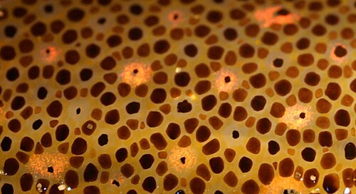
Key Facts
- Chromatophores are pigment-containing cells found in many ectothermic animals.
- Color change through pigment translocation is known as metachrosis.
- Cephalopods use muscular control for rapid color change, while vertebrates rely on cell signaling.
- Melanophores are responsible for black and brown tones using the pigment eumelanin.
- Defects in melanin synthesis can lead to conditions such as albinism.
Classification of Chromatophore Types
Chromatophores are categorized into several subclasses based on the specific colors they produce under white light. While most rely on pigments that absorb certain wavelengths, some utilize light scattering and optical interference to create color.
Pigment-Based Cells: Xanthophores and Erythrophores
The distinction between yellow and red cells can sometimes be fluid. Xanthophores contain large amounts of yellow pteridine pigments, while erythrophores primarily contain red or orange carotenoids. Because vesicles containing both types of pigments can exist within a single cell, the resulting color depends on the specific ratio of red to yellow pigments present.
Structural Color: Iridophores and Leucophores
Unlike pigment-based cells, iridophores and leucophores produce color through physical properties. Iridophores create reflective or iridescent effects, while leucophores appear white. These colors are generated by the way these cells scatter light or utilize optical interference.

The Role of Cyanophores
In addition to the common types, some species possess cyanophores, which are specialized for producing blue coloration.
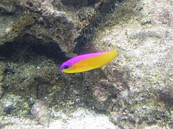
Melanophores and the Science of Dark Pigmentation
Melanophores are perhaps the most widely studied chromatophores. They contain eumelanin, a dark brown or black pigment produced from the amino acid tyrosine. This pigment is packaged into vesicles called melanosomes and distributed throughout the cell.
The synthesis of melanin is driven by the enzyme tyrosinase. If this protein is defective, the organism cannot generate melanin, resulting in albinism. Interestingly, some species exhibit more complex pigment profiles. For example, certain phyllomedusine frogs possess a deep wine-red pigment within their melanophores, and some anole lizards use a pteridine dimer called pterorhodin to achieve colors ranging from bright blue to black.
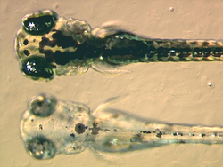
In fish and frogs, melanophores can change color by dispersing or aggregating these pigment-containing bodies. This movement is regulated by various receptors, including a homologue of the human MC1R receptor, which is essential for melanin dispersion in zebrafish.
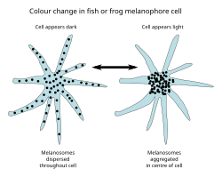
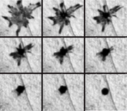
Mechanisms of Rapid Color Change
The ability to change color rapidly—a process called physiological color change or metachrosis—is a vital survival tool. This occurs through two primary methods:
- Pigment Translocation: Inside the cell, molecules like cyclic adenosine monophosphate (cAMP) act as messengers, driving molecular motors to move pigment vesicles along microtubules and microfilaments.
- Structural Reorientation: Some animals reorient reflective plates within their cells to alter light reflection.
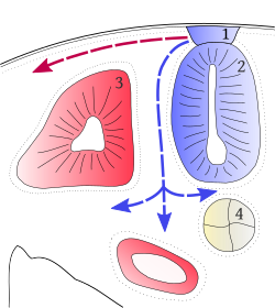
The control mechanisms differ significantly between groups. Cephalopods, such as the octopus, possess complex chromatophore organs controlled directly by muscles. In contrast, vertebrates like chameleons achieve color changes through cell signaling involving hormones or neurotransmitters, often triggered by stress, temperature, or environmental changes.
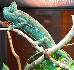
Development and Background Adaptation
Chromatophores develop from multipotent precursor cells called chromatoblasts. In zebrafish embryos, all major classes of chromatophores are present by three days after fertilization. Research into transcription factors like kit, sox10, and mitf has shown that if these proteins are defective, chromatophores may be absent, leading to a leucistic disorder.
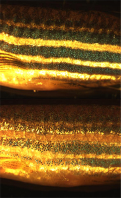
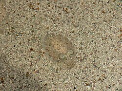
Many species also exhibit background adaptation, where they adjust their coloration to match their surroundings. This is seen in zebrafish, which adapt to light or dark environments, and in infant cuttlefish, which use these mechanisms to mimic their local environment.
Summary of Chromatophore Types
| Type | Primary Color | Mechanism |
|---|---|---|
| Xanthophore | Yellow | Pteridine pigments |
| Erythrophore | Red / Orange | Carotenoid pigments |
| Melanophore | Black / Brown | Eumelanin pigments |
| Iridophore | Iridescent | Light reflection/interference |
| Leucophore | White | Light scattering |
| Cyanophore | Blue | Pigment-based |
Frequently Asked Questions
How do cephalopods change color so quickly?
Unlike vertebrates that rely on slower hormonal signaling, cephalopods like octopuses have complex chromatophore organs that are controlled directly by muscles, allowing for near-instantaneous color shifts.
What is the difference between a chromatophore and a melanocyte?
Chromatophores are the primary color-producing cells in many ectothermic animals (like fish and reptiles). Melanocytes are a specific class of cells used by mammals and birds for coloration.
What causes albinism in animals?
Albinism can occur when the enzyme tyrosinase is defective, preventing the synthesis of melanin within the melanophores.
Can animals change color due to their mood?
Yes, in vertebrates like chameleons, color change can be initiated by neurotransmitters or hormones in response to changes in mood, stress, or temperature.
Why are zebrafish used in chromatophore research?
Zebrafish are valuable model systems because their chromatophore development is well-understood, and they allow scientists to study how cells organize into complex patterns and communicate.