Bone Biology: Structure, Composition, and Clinical Health
Bone is a specialized living tissue that serves as the structural framework of the body. Far from being static, bones are dynamic organs that constantly remodel themselves to adapt to mechanical stress and maintain mineral homeostasis. From protecting vital organs to enabling movement, the skeletal system is a masterpiece of biological engineering.

Key Facts
- Composition: Bone consists of approximately 70% inorganic minerals and 30% organic matter.
- Primary Mineral: Hydroxyapatite is the dominant inorganic component, providing compressive strength.
- Organic Matrix: Type I collagen provides the necessary tensile strength and elasticity.
- Mechanical Strength: Bone is strongest under compression (170 MPa) and weakest under shear stress (51.6 MPa).
- Cellular Activity: Bone health is maintained by a balance of osteoblasts (bone-forming cells) and osteoclasts (bone-resorbing cells).
Gross Anatomy and Mechanical Properties
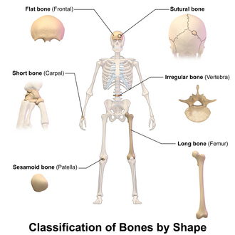
Bones can be classified by their shape and appearance, which typically dictates their function within the body.
The mechanical integrity of bone is defined by its ability to handle different types of stress. Bone possesses a high compressive strength (about 170 MPa), meaning it resists pushing forces exceptionally well. However, it has lower tensile strength (104–121 MPa) and very low shear stress strength (51.6 MPa), making it susceptible to torsional loads. While bone is generally brittle, the presence of collagen provides a significant degree of elasticity.
Composition and Histology
At the microscopic level, bone is a composite material consisting of living cells embedded in a mineralized organic matrix.
The Inorganic Phase
Roughly 70% of bone mass is inorganic. The primary component is hydroxyapatite, a mineral with the nominal composition Ca10(PO4)6(OH)2. This mineral phase, along with other calcium and phosphate salts, provides the bone with its rigidity and compressive strength. The ratio of calcium to phosphate typically varies between 1.3 and 2.0 by weight, with trace amounts of sodium, potassium, magnesium, and carbonate.
The Organic Phase
The remaining 30% of the acellular component is organic, consisting mainly of type I collagen. These collagen fibers act as a reinforcement, providing tensile strength and preventing the bone from being overly brittle. The synergy between the flexible collagen and the hard hydroxyapatite crystals allows bone to withstand complex physical loads.
Bone Cells
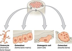
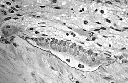
Three primary cell types manage the lifecycle of bone tissue:
- Osteoblasts: Cells responsible for synthesizing the organic matrix (osteoid) and initiating mineralization.
- Osteocytes: Mature bone cells trapped within the matrix that monitor and maintain the tissue.
- Osteoclasts: Large cells that break down and resorb bone tissue during remodeling.
Bone Development and Remodeling
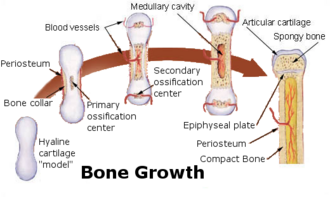
Bone formation occurs through various processes, most notably endochondral ossification, where cartilage is gradually converted into bone tissue. This process is critical during fetal development and continues at the growth plates in children.
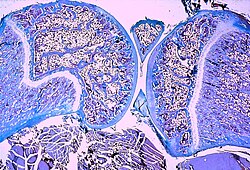
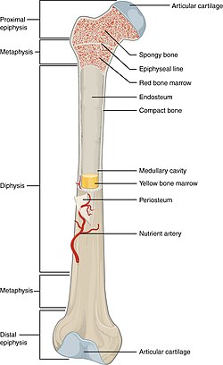
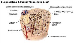
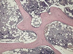
Once formed, bone undergoes constant remodeling. This process ensures that old or damaged bone is replaced and that the skeleton can adapt to new physical demands. This is often seen in the transition between cortex (the dense outer shell) and trabeculae (the spongy, lattice-like inner bone).
Clinical Significance and Bone Health
Various conditions can compromise the structural integrity of the skeleton, leading to increased fragility.
Fractures and Density
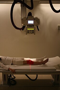
Fractures occur when the stress applied to a bone exceeds its mechanical strength. Radiography is the primary tool used to identify these breaks.
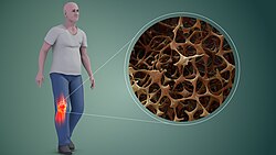
Osteoporosis is a condition characterized by reduced bone mineral density (BMD), which significantly increases the likelihood of fractures due to the thinning of the bone matrix.
The Impact of Diabetes
Recent research suggests a strong link between diabetes and bone health. Both Type 1 and Type 2 diabetes may inhibit osteoblastic activity, leading to lower bone mineral content (BMC) and BMD in both children and adults. This weakening increases the risk of osteopenia, osteoporosis, and osteoarthritis, potentially limiting athletic performance and daily activities.
Other Conditions
- Osteogenesis imperfecta: A genetic disorder affecting collagen production.
- Ankylosing spondylitis: An inflammatory disease affecting the spine.
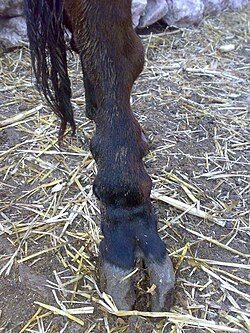
- Skeletal fluorosis: Bone deformation caused by excessive fluoride intake, often due to industrial contamination.
Comparative Osteology
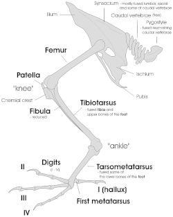
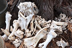
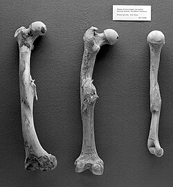
Bone structure varies across species to meet specific evolutionary needs. For example, birds possess lightweight skeletons to facilitate flight, while aquatic mammals may develop denser bones for buoyancy control.
Summary of Bone Properties
| Property | Details | Primary Component |
|---|---|---|
| Inorganic Phase | ~70% of mass; provides compressive strength | Hydroxyapatite |
| Organic Phase | ~30% of mass; provides tensile strength | Type I Collagen |
| Compressive Strength | ~170 MPa | Mineral crystals |
| Shear Strength | ~51.6 MPa | N/A (Weakest point) |
| Key Cells | Formation, Maintenance, Resorption | Osteoblasts, Osteocytes, Osteoclasts |
Frequently Asked Questions
What is the difference between osteoblasts and osteoclasts?
Osteoblasts are the cells responsible for building new bone by synthesizing the organic matrix and aiding mineralization. In contrast, osteoclasts are cells that break down and resorb bone tissue, allowing for remodeling and the release of minerals into the bloodstream.
Why is collagen important for bone strength?
While minerals make bone hard, collagen provides flexibility and tensile strength. Without collagen, bones would be extremely brittle and would shatter easily under tension or impact.
How does diabetes affect bone health?
Diabetes (both Type 1 and Type 2) is thought to inhibit the activity of osteoblasts. This leads to a decrease in bone mineral density and content, which increases the risk of developing osteoporosis and suffering fractures.
What is hydroxyapatite?
Hydroxyapatite is the primary inorganic mineral found in human bone, consisting of calcium and phosphate. It forms crystals that intersperse with collagen fibers to give bone its characteristic hardness and compressive strength.
What is endochondral ossification?
Endochondral ossification is the biological process by which a cartilage template is replaced by bone tissue. This is the primary method by which long bones grow in length during childhood.